Dermoscopy or dermatoscopy refers to the examination of the skin using skin surface microscopy, and is also called ‘epiluminoscopy’ and ‘epiluminescent microscopy’. Derm(at)oscopy is mainly used to evaluate pigmented skin lesions. In experienced hands it can make it easier to diagnose melanoma.
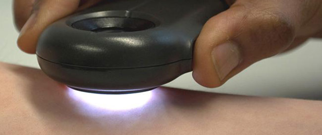
Dermatoscopy requires a high quality magnifying lens and a powerful lighting system. This allows examination of skin structures and patterns. There are several different lightweight, battery-powered hand-held devices. Convenient attachments allow video or still photography.
Computer software can be used to archive dermatoscopy images and allow expert diagnosis and reporting (mole mapping). Smart programs may aid in diagnosis by comparing the new image with stored cases with typical features of benign and malignant pigmented skin lesions.
Dermatoscopic features of pigmented lesions
Using dermoscopy, the pigmentation of the lesion is evaluated in terms of colour(s) and structure. Colours found in pigmented skin lesions include black, brown, red, blue, grey, yellow and white.
Characteristics of the dermatoscopic structure of the skin lesions include:
- Symmetry or asymmetry
- Homogeny/uniformity (sameness) or heterogeny (structural differences across the lesion)
- Distribution of pigment: brown lines, dots, clods and structureless areas
- Skin surface keratin: small white cysts, crypts, fissures
- Vascular morphology and pattern: regular or irregular
- Border of the lesion: fading, sharply cut off or radial streaks
- Presence of ulceration